Is Ultrasound Imaging Safe? A Simple Breakdown
Over the years, a recurring question that lands in my inbox and on my phone line is: “Is ultrasound safe?” It’s a valid concern, especially in an age where information is abundant, yet clarity can be scarce. Many expectant mothers express hesitance towards undergoing ultrasound scans, often rooted in the uncertainty of long-term data about its safety. Many people are now waking up and starting to question the mainstream narrative. This can be a scary place to be on your journey especially when one comes to question the validity in everything we have been taught.
While I firmly believe in empowering individuals to make informed decisions, I also understand the apprehension that can arise when confronted with dense scientific studies. They can be daunting, overflowing with jargon, and difficult to decipher without a background in the field.
That’s why I’ve decided to bridge that gap. One study that I frequently recommend has now been condensed into this blog post. My aim? To present its findings in a more digestible and comprehensible manner, without the technical overload. This way, you can gain a clearer understanding and make the decision that’s right for you and your baby.
Remember, knowledge is power, but understanding is the key. And if you ever find yourself needing further clarification or support, please don’t hesitate to reach out. Your peace of mind is paramount.

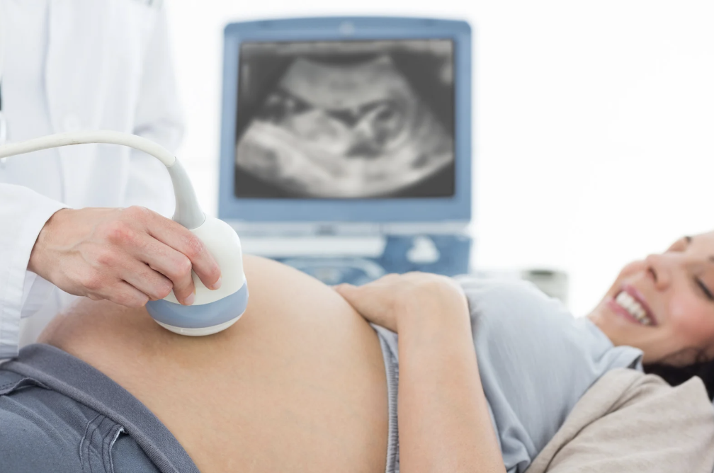
Ultrasound imaging is a tool doctors use for various reasons:
Pregnancy: Checking on babies before they’re born.
Cancer: Observing tumours and seeing if treatments are working.
Heart Health: Examining how the heart is functioning.
Recently, some places even offer parents a chance to get a “first photo or video” of their unborn baby, outside of regular medical checks.
But is it safe? As the popularity of ultrasound grows, it’s crucial to understand any risks. This article dives into research on the topic to provide some answers.
How Does Ultrasound Work and Is It Safe?
When we talk about ultrasounds, we often hear the term “non-invasive.” This means that it doesn’t require any cuts or contact with our insides. However, it’s important to understand that for ultrasound to create an image, it sends ultrasonic energy (or sound waves) into the body.
Here’s a simple breakdown of how it works:
- Sending the Sound Waves: The ultrasound machine sends out sound waves into the body.
- Energy Behaviour: As these waves travel deeper, some of the energy bounces back, while some get absorbed. Things like bones reflect more energy than soft parts, which is why they appear brighter on the ultrasound image.
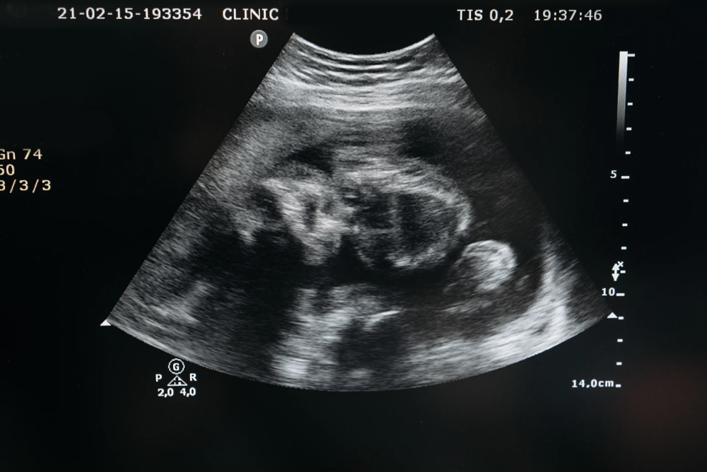
- Creating the Image: The machine reads the bounced-back energy to produce an image. How deep this energy comes from and how strong it is, helps in understanding what’s inside.
Now, when it comes to safety, there are two main ways ultrasounds can affect tissues:
- Thermal Effects: This is about heating. When tissues absorb the sound wave energy, they can warm up. The amount of heating can vary based on the type of ultrasound and what it’s looking at. For instance, bones absorb more energy and can get warmer than softer tissues. The most heat is absorbed when the ultrasound is near bones or tissues close to bones, especially in growing bones.
- Non-Thermal Effects: Apart from heating, there are other ways ultrasounds can interact with tissues, but they’re often closely linked with thermal effects. For example, heating can make non-thermal effects more likely, and vice versa.
In simple terms, the main concern is the heating, especially near bones. But, many factors, like the movement of the device and blood flow, can cool the area down and make the actual heating less than what’s estimated.
How Much Does Ultrasound Heat Our Body
Ultrasound can heat up parts of our body as it sends its sound waves. Here’s what you need to know:
Cooling Effects: Not every part of our body heats up the same. Organs with a lot of blood, like the liver or kidney, cool down faster. That’s because the blood flow can cool the heated areas. But in some types of ultrasound, the heat might not spread out quickly enough, causing higher temperatures in small spots.
Duration Matters: How hot something gets isn’t the only concern – how long it stays hot is also important. There’s a concept called ‘thermal dose’. In simple terms, this refers to how long a specific body part is exposed to a certain temperature. Shorter exposures to higher temperatures can have similar effects as longer exposures to lower temperatures.
Effects of Heat: Too much heat can be harmful, especially for unborn babies. There have been studies on the effects of heat on embryos. The main concern is the brain of the developing baby. How much heat can cause harm often depends on the stage of the baby’s development. While some stages can handle more heat, others, like right after conception, are very sensitive. Most studies focus on heating the whole body, which is different from the focused heating from an ultrasound. However, during late pregnancy, the bones in the baby can get hotter than earlier stages.
Safe Temperature Limits: There’s debate on how hot is too hot. Some experts suggest it’s okay if our body’s temperature rises by 1-1.5°C for a long period. Others say a rise of up to 2.5°C for over an hour is the limit. For very high temperatures, the safe limit is short – like a rise of 4°C for only 30 seconds.
How Do We Know? It’s tough to know exactly how hot tissues get during an ultrasound. There’s a tool called the ‘thermal index’ that gives an estimate, but it’s not always precise.
Non-Thermal Effects of Ultrasound: What Happens Besides Heating?
Ultrasound does more than just heat tissues. There are also mechanical effects:
Acoustic Streaming: This is the main safety concern. It refers to how the ultrasonic waves move through tissue.
Bubbles and Cavitation:
Bubble Formation: When ultrasound waves pass through tissues, the negative pressure can cause gas bubbles to form. This process, related to the formation of bubbles in deep-sea divers during rapid ascension, is called acoustic cavitation.
Bubble Behaviour: Once formed, these bubbles can expand and contract. There are two main types:
Stable Cavitation: Bubbles oscillate without breaking up. This can cause small currents in fluids and might change how cells transport ions, potentially having therapeutic benefits.
Inertial Cavitation: Bubbles grow rapidly and then collapse, leading to high temperatures and pressures that can damage tissues.
Bubble Size and Resonance: The size of a resonant bubble changes with the frequency of ultrasound. For frequencies between 1 and 3 MHz, the bubble size is around 4-1 µm.
Debate on Cavitation: There’s some disagreement on whether typical ultrasound can cause cavitation without external bubbles (like those from contrast agents). Most experts think it’s unlikely, but not impossible. Cavitation can be detected by special sensors that listen for the sounds of the bubbles.
Radiation Pressure & Acoustic Streaming:
Ultrasound can exert force on things in its path due to radiation pressure and can cause fluid movement, known as acoustic streaming.
High velocities and stresses may be produced, especially near boundaries. For example, measurements have shown velocities up to 14 cm/s during certain ultrasound modes.
Research Limitations: Much of the available research on ultrasound effects doesn’t use conditions similar to medical scenarios.
Cell experiments are valuable but might show different effects due to the water environment, promoting cavitation.
Experiments on tissues, whether inside or outside a body, can provide more accurate conditions, but still have their limitations.
Most safety studies use small animals, which have different proportions of exposure than humans.
In essence, while ultrasound has potential non-thermal effects, the exact consequences and risks are not fully clear.
MACHINE OUTPUTS
The Challenge: Understanding the effects of ultrasound from studies can be hard because we need to compare the settings used in those studies to what’s actually used in hospitals and clinics.
How Machines Work: Ultrasound machines send out sound waves, and they measure them in two main ways:
- The total power of the sound (like volume on a speaker).
- The pressure of the sound wave at different points.
Reading Good Research: The best research articles give full details on how the ultrasound was used, like its volume, how long it was on, and at what frequency.
Misunderstandings: In X-rays, there’s a difference between exposure (how much you’re exposed to) and dose (how much actually enters and affects your body). But with ultrasound, people sometimes use those terms like they mean the same thing, which can be confusing.
Real World vs. Lab: It’s hard to measure the exact intensity of ultrasound inside the body, so researchers often estimate it based on what they know about the tissues it’s passing through.
What Manufacturers Say: A recent survey looked at the power levels of ultrasound machines from big manufacturers. They found that these power levels have generally gone up over the years, but it varies depending on the mode the machine is in.
ANIMAL STUDIES
Lots of Research: There’s been a lot of research on how ultrasound affects animals, from single cells to whole animals.
Lab Setups:
- Tests on cells in dishes can show how ultrasound affects cells directly, but it’s not the same as in a living body.
- Tests on whole animals often use stronger ultrasound settings than what’s used in human check-ups.
What We’ve Seen:
- Ultrasound can cause tiny bleeds in animal lungs and guts, but only in parts with air (like gas pockets). The effects of these tiny bleeds are still uncertain.
- For baby animals in the womb, most ultrasound tests don’t show any harm. However, a couple of studies did show changes like lower birth weights in monkeys and affected brain cell movements in mice. But it’s tricky to know if these findings would apply to humans.
Learning and Memory: One study on baby chick brains showed that a certain type of ultrasound might affect learning and memory, but it’s unclear if this would be the same for humans.
The Takeaway: In a lab setting, very strong ultrasound can break cells apart, especially in water. In animals, there’s a slight chance that ultrasound could affect certain tissues, especially if they’re developing or have gas in them. For baby animals in the womb, the stage of their development seems to matter in how they react to ultrasound. Some results might be concerning, but we’re still figuring out what they mean for humans.
UNDERSTANDING ULTRASOUND SAFETY IN PREGNANCY
Ultrasound has been used for many years during pregnancy to check on the baby’s health. While it seems to be safe, scientists like to be extra sure, which is why they’ve done a lot of studies to check its effects.
So far, most studies show that ultrasounds haven’t caused any obvious harm to babies before they’re born. However, absence of evidence of harm is not evidence of absence of harm!
Researchers have looked at various things that might be affected by ultrasound, like the baby’s weight at birth, their health right after birth, and even how they do in school. Most of these studies show that ultrasound doesn’t seem to cause any problems.
One interesting finding is that boys who had ultrasounds when they were still in the womb might be more likely to be left-handed. But being left-handed isn’t a bad thing, so scientists are just curious about why this might be the case.
One important thing to note is that many of the older studies used older ultrasound machines. The newer machines are a bit stronger. So, while the track record looks good, doctors and technicians need to stay informed and careful.
Diagnostic ultrasound uses pulsed sound waves. A study quoted in the New Scientist indicated that an ultrasound emits a sound similar to the highest piano notes, reaching up to 100 decibels, comparable to a subway train. However, the frequencies used in diagnostic ultrasound (1 – 10 megahertz) are beyond human audible range. While some reports have indicated the potential of babies to hear certain ultrasound frequencies, others have disproven this notion. Additionally, reports on potential harm to babies from ambient noise, like from industrial environments, differ, with some studies showing no adverse effects.
2010 Clinical Safety Statement
Below is a summary overview from the 2010 Clinical Safety Statement on Diagnostic Ultrasound. You can find the full report here
What is Diagnostic Ultrasound?
It’s a tool doctors use to see inside your body, often used during pregnancy to look at the baby. It has been used for a long time without causing harm. But, like all tools, if not used correctly, it could be harmful.
Why the Concern Now?
Even though it’s safe, doctors are using it more frequently, in more ways, and with stronger machines. So, we need to be extra careful.
Who Should Use It?
Only trained professionals who know how to use it safely. The machines also need to be in good condition.
How Does Ultrasound Work and What are the Concerns?
Ultrasound sends waves into the body, which can create heat and pressure. Too much heat can be dangerous, especially for unborn babies. There have been some concerns from animal tests, but we haven’t seen these issues in humans unless they were using a special agent that helps with the ultrasound.
What’s on the Screen When Using Ultrasound?
The machine shows two main indicators:
Thermal Index (TI): Tells the user about potential heating in the body.
Mechanical Index (MI): Indicates possible non-heating effects.
Doctors should keep these values as low as possible and still get a clear image. If the values are too high, they should use the machine for a shorter time.
Are There Specific Modes to Watch Out For?
Yes, certain settings, like Doppler and colour imaging, can produce more heat. There’s also 3D and 4D imaging, with 4D being like a live video. 4D can mean continuous exposure, so doctors shouldn’t use it longer than needed.
What About Ultrasound During Pregnancy?
Unborn babies are delicate, especially in the early stages. Doctors should be extra careful, using the lowest settings and for the shortest time to get the needed information. Bones in the baby can get very warm from ultrasound, especially as they harden, so caution is needed there. While ultrasounds are safe for pregnancies when needed, Doppler ultrasounds (a specific kind) shouldn’t be used in the first three months of pregnancy.
Are There Other Sensitive Areas?
Yes, when checking the eyes or the heart and head of newborns, extra caution is required.
What About Ultrasound Enhancing Agents?
There are agents that can be used to make ultrasounds clearer. But, these agents can sometimes cause side effects, especially in small blood vessels. This is a concern in the brain, eyes, and for newborns. So, doctors should be very careful when using them.
Are newer ultrasounds safe?
We don’t have conclusive evidence about the safety of today’s ultrasound machines. The majority of the safety information we have comes from older machines used before the mid-1990s. Newer devices have much stronger output levels. Therefore, it’s uncertain if current studies can guide us about the safety of present-day ultrasounds.
Ultrasound operators and safety information:
There’s a feature on ultrasound machines that shows safety data in real-time. This was introduced in the 1990s. However, a study found that many professionals using ultrasound weren’t familiar with these safety features. Basically, many operators didn’t know how to adjust or monitor the ultrasound’s energy levels, which is concerning.
Concerns about using Doppler ultrasound early in pregnancy:
There’s a specific type of ultrasound called Doppler. Experts have advised caution when using it in early pregnancy. However, its use has grown as it can help assess the risk of certain conditions. The key concern isn’t that we know it’s harmful, but that we don’t have enough information to confirm it’s completely safe, especially during the first trimester when the baby is particularly sensitive.
Ultrasound research and safety:
Back in 1999, a medical journal set strict guidelines for publishing research involving Doppler ultrasounds in the first trimester. But a quick look at recent papers suggests that not all studies are strictly following these guidelines. This might indicate a need to revisit and emphasise these safety guidelines in medical research.
In short, while ultrasounds have been a valuable tool in prenatal care, it’s important to be aware and cautious of potential safety issues, especially with newer devices and specific techniques like Doppler during early pregnancy.
Conclusion
In the world of medical advancements, uncertainties are inevitable. When it comes to ultrasound safety, it’s no different. Just because we haven’t found evidence of harm doesn’t mean it’s entirely without risk. Every medical procedure has its risks, and the absence of proven danger doesn’t guarantee complete safety.
In the vast realm of ultrasounds and their intricacies, I’m no expert. But as I pen down my thoughts on this blog, it’s challenging to encapsulate the entire breadth of knowledge available on this topic. While I’ve tried to be as comprehensive as possible, there’s a good chance I might have overlooked some pivotal information or recent studies. Thus, I warmly invite feedback from all you informed readers out there. If there’s any essential piece of evidence or study that I might have missed, please do not hesitate to share in the comments below.
The old adage goes, “It takes a village.” And in the age of information, pooling our collective wisdom can be a game-changer. When we come together and share our knowledge, we can better guide those seeking insights. This is especially true when it comes to making informed choices about one’s health and wellbeing.
A crucial aspect I’d like to emphasise is the importance of heeding one’s intuition, especially for women. Far too often, we’re conditioned to doubt our instincts, to always look outside for answers. But within each of us lies a deep, intuitive wisdom – a gut instinct that should never be ignored. So, to all the women reading this, if something doesn’t sit right with you or raises questions, trust yourself. Listen to that inner voice.
For those who have a burning passion to delve deeper into the intricacies of childbirth or are contemplating launching a career as a professional birth keeper, I highly recommend our comprehensive one-year birth keeper course. This program isn’t just another certification; it’s a journey that covers a multitude of topics ranging from trauma-informed care, human rights in childbirth, medical ethics, to specialised areas like breech birth, homoeopathy, and even spiritual development during childbirth.
Our course isn’t just about imparting knowledge; it’s about empowering and equipping individuals to be a beacon of change in the realm of childbirth. We believe in leading a revolution where each birth becomes an empowering experience. The ultimate goal? To change the narrative of childbirth, one birth at a time, and to bolster women’s confidence in their choices and instincts.
Let’s rally together to serve women and put their needs and instincts at the forefront, instead of merely conforming to a system. Join us in this transformative journey to usher in a new era of empowered childbirth.
References
https://obgyn.onlinelibrary.wiley.com/doi/full/10.1002/uog.6381


